买球有限公司")
Successful case
Our advantage
买球入口登录官网(中国)买球有限公司
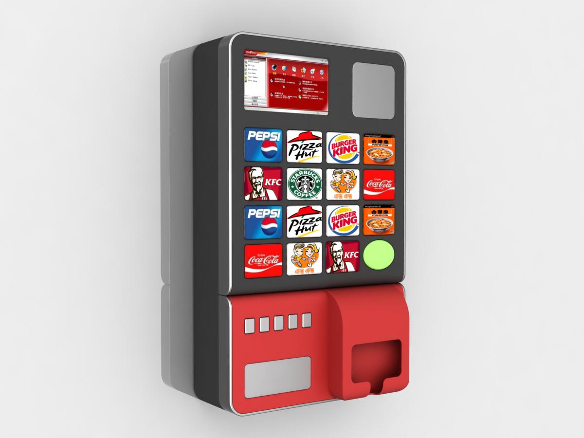
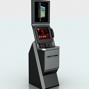
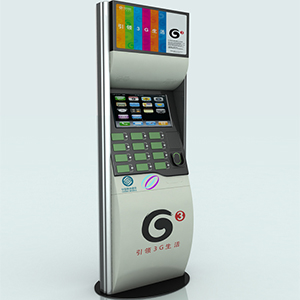
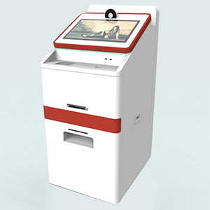
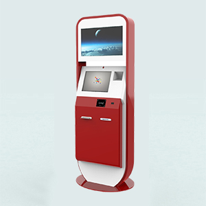
买球入口登录官网(中国)买球有限公司(BNFUTURE)成立于1998年,主要从事触摸屏自助终端的设计和研发。研发的产品种类丰富,包括门禁访客自助机、优惠卷打印自助设备、信息亭自助机、民航自助值机等产品。行业涉及金融、电信、交通、工商、税务、医疗、房地产、政府机关、大型企业等。拥有专业的工业设计团队,具备提供应用行业系统硬件的解决方案能力,专注于工业产品在现代信息领域的应用与发展。
北纳科技经历多年发展,逐步形成了一套完整设计研发体系并不断完善。把工业设计创新作为公司日益发展的源泉,以超越自我的动力,不断以产品的创新和应用方案的解决来满足市场和用户的需求。
北纳科技把质量管理看作企业发展的生命线,并一直将"用户的满意"作为自己的质量标准。目前,公司的质量保证体系己覆盖了影响产品和服务质量的所有环节,包括质量管理、产品生命周期各阶段、产品支持活动、生产供应物流系统的各个阶段,使产品和服务的全过程纳入规范化,程序化的管理和控制之下。
本公司采用规范的现代化管理制度、管理结构及管理体制,并汇集了一批自助领域的专业人才,形成了一支志同道合的精英团队。
北纳科技将砺精图志,不懈努力,为推动中国工业设计的进步作出一份贡献。